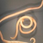
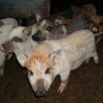
Trichinosis is an invasive disease of many animals and humans. It is caused by Trichinella - small round worms related to nematodes, parasitic only inside the host's body. Trichinosis in domestic pigs is especially dangerous. If sanitary standards are neglected when slaughtering animals and processing their meat, parasite larvae can enter the human body. Trichinosis is dangerous because there are still no reliable treatments for farm animals.
How does the parasite develop?
The development of the pathogen occurs inside the body in several stages.
Intestinal phase
The parasite enters the stomach of a pig or other animal in the form of live encapsulated larvae along with meat food. There, the capsules are destroyed by gastric juice, and the larvae end up in the duodenum. There they stay for 30-40 hours. During this time, they form into adults capable of reproduction. Fertilization occurs. The males die after it.
Fertilized females insert their head end between the tubular outgrowths and villi of the intestinal epithelium. The development cycle from egg to larva occurs inside it within 7 days. A week later, females give birth to live larvae. After this, the next phase of development begins.
One individual can produce about 2000 larvae. Trichinella that lay larvae remain alive for up to 8 weeks, after which they die and are excreted along with feces.
Migration phase
The migration routes of the larvae are still the subject of scientific controversy. It is believed that they first enter the lymph. From there, the pathogen migrates to the lymph nodes, from which to the vena cava. Then they spread through the bloodstream throughout the pig’s body. The size of migratory larvae does not exceed 110 microns in length and 5-6 microns in diameter. Their movement continues until they enter the skeletal muscle tissue.
Muscle phase
Inside the striated muscle, the larvae are trapped under the cell membranes. The muscle larva has the following dimensions:
- males are 1.1 mm long and 0.06 mm in diameter;
- females are 1.3 mm long and 0.06 mm in diameter.
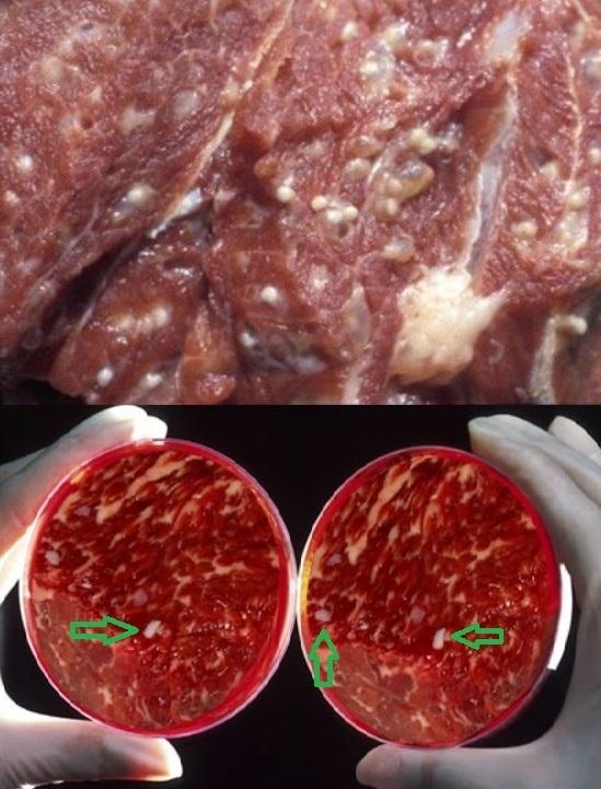
In the larvae, during the muscular stage, the main organs are formed. They increase significantly in size. The length of females is 3-4, and males - 2.2 millimeters.The larvae curl into a spiral and a capsule appears around them. Its formation takes from 3 to 9 weeks. It will take approximately another 16 months for calcification. In skeletal muscle, encapsulated larvae can remain alive for up to 25 years. In the external environment, this period is equal to six months.
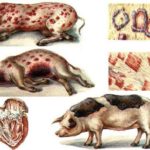
Symptoms of trichinosis
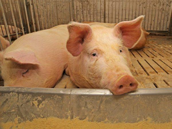
In case of accidental infection of pigs, the clinical picture is blurred. In approximately 30% of cases there are no symptoms. With intensive intentional infection in animals, damage to the intestines and blood vessels is observed. The disease is characterized by the following symptoms:
- refusal to eat;
- lethargic behavior;
- weight loss;
- exhaustion;
- convulsions;
- swelling;
- vomit;
- diarrhea;
- breathing problems;
- allergic manifestations in the form of a rash.
During life, the disease can be diagnosed in a domestic pig by a combination of symptoms, analysis of tissue taken from the ears, or through a special enzyme-linked immunosorbent assay. Wild boar meat is most often subjected to post-mortem examination.
Rules for diagnosing the disease
To confirm the well-being of pig herds, intravital diagnostics of animals is of great importance. ELISA is of great importance in this regard. This is a special enzyme immunoassay. It is carried out on animals of any age, starting from a full 3 months of life, 3-4 weeks after infection. Identified sick individuals are excluded from the fattening group. The disadvantage of such a study is considered to be 92-97% accuracy. Therefore, ELISA does not exclude further examination of pig carcasses. To confirm the results and more accurately diagnose, a veterinary examination is carried out in several ways. For this use:
- Compressor trichinoscopy, for which 24 sections are made from muscle tissue, crushed, and then examined under a trichinelloscope or microscope.
- Digestion of muscles in artificial gastric juice. A very accurate method of post-mortem diagnosis. The selected meat is ground and poured with a mixture of water, hydrochloric acid, and pepsin. Place it in the thermostat for 5 hours. Then the liquid is drained and the sediment is examined.
- Post-mortem diagnosis is carried out with a complete autopsy of the intestine. Adult parasites are most often found in the small intestine. A section of the intestine is cut and filled with water along with its contents. After several washes, the sediment is examined under a microscope.
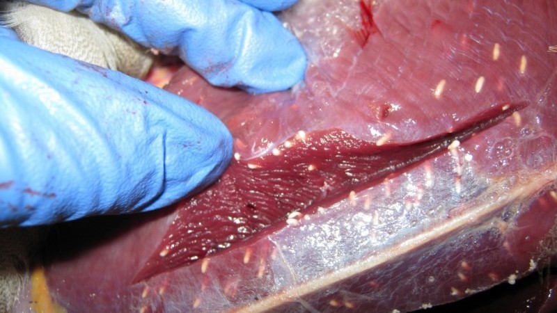
These methods make it possible to identify Trichinella in raw meat carcasses and ready-to-eat meat products. Currently, IFR is becoming popular - an enzyme-linked immunosorbent reaction that is carried out on special polymer membranes.
Treatment methods for porcine trichinosis
Modern veterinary pharmacology has not developed effective drugs that can treat pigs for trichinosis. The following drugs can be used:
- Thiabendazole;
- Mebendazole;
- Albendazole;
- Parbendazole.
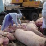
These agents are effective only against adults and larvae up to the capsule formation stage. There is information about treatment with cadmium oxide and chlorophos. In most cases, animals infected with Trichinella are euthanized. The carcasses are disposed of.
Why are parasites dangerous?
Trichinella larvae enclosed in capsules are dangerous because they are resistant to high and low temperatures and other destructive processes. When cooking a piece of meat weighing about 1 kg, the destruction of the larvae can be achieved only after two and a half hours. The freezing process at -25 degrees must be carried out for at least 4-5 days. In rotting remains, the larvae remain alive for 4-6 months.
Parasites are very dangerous for humans.Poorly cooked, undercooked meat from infected pigs can become a source of infection. Complete recovery from trichinosis can occur after 6-12 months of complex therapy. In severe cases, it can cause various serious disturbances in the functioning of the heart and central nervous system, which causes the death of the patient.
Prevention measures
To prevent trichinosis, it is necessary to protect areas where pigs are kept from rodents. Rats and mice are carriers of this disease. Strong walls, floors, and the lack of holes in them will become an obstacle for them.
It is advisable to bury the carcasses of animals obtained during hunting to a depth of at least a meter, in places inaccessible to tearing by pigs, dogs, and other animals. Do not feed raw residues from the slaughter of domestic animals and fur-bearing animals to pigs. Follow the rules for slaughtering livestock on private farms and farms. To prevent infection of people, you should not purchase pork that has not passed a veterinary examination. Meat obtained from hunting must be taken to the nearest laboratory for testing. It is important to remember that any disease is easier to prevent than to treat.