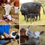
For cattle owners, knowledge about the structure of the cow's udder and the diseases to which the organ is susceptible is necessary. They allow you to properly care for livestock, maintain animal productivity, and promptly respond to problems that arise. Proper milking of first-calf heifers, massage, and systematic inspections guarantee the health of the dairy herd and the quality of the resulting products.
How does a cow's udder work?
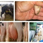
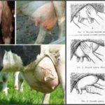
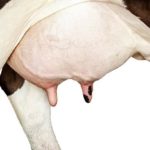
The udder of cattle is located in the groin area.The organ is covered with delicate sparse hairs; in the rear part the hairs grow from bottom to top and to the sides and form a “milk mirror”, by the size of which one can judge the productivity of the animal. The structure of the udder is complex; the milk production of an animal depends on the interaction of many systems: digestive, hormonal, and the central nervous system (CNS).
The udder consists of glandular tissue - parenchyma, adipose and connective tissue. The glandular tissue has many alveoli - vesicles in which milk production occurs; adipose and connective tissue protect the parenchyma from external influences (hypothermia, overheating, bruises and injuries). These tissues are penetrated by blood vessels. In highly productive animals, blood vessels are clearly visible under the skin; the amount of milk produced depends on the degree of blood supply to the udder.
Under the influence of hormones (oxytocin, prolactin, estrogens), the alveoli begin to produce milk. It enters small ducts extending from the alveoli. Small ducts unite to form medium ducts, which, in turn, “flow” into 12-50 large milk ducts leading down to the milk tanks of the nipple. The milk cistern is a cavity in the upper part of the nipple, connected to the parenchyma, in which milk accumulates.
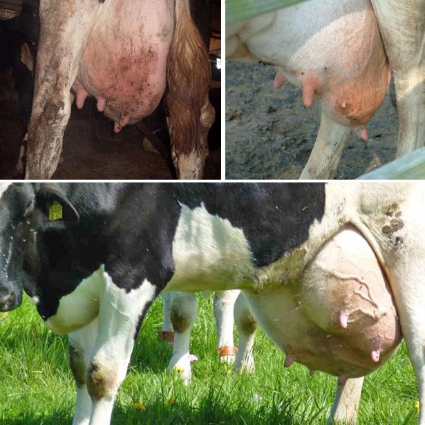
The mammary gland consists of 4 lobes, each of which ends in a nipple. The shares are a closed system for milk production; they are not connected to each other. The right and left lobes of the udder are separated by an elastic septum - a ligament that supports the organ. The posterior lobes are more developed than the anterior ones. Cows that produce a lot of milk have well-developed nipples, they are located far apart and are 8-10 centimeters long.The nipple consists of a base that passes into the body of the lobe, an apex (lower part) and a cylindrical middle part.
The nipples have many nerve endings; during milking, they become irritated and send signals to the animal’s brain, causing milk to be released. The walls of the nipple are lined with muscle fibers, which form a sphincter at the top of the nipple - a locking device that prevents the free flow of milk.
Development
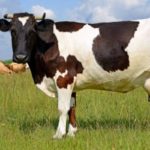
The udder of a heifer is laid during fetal development. The productivity of an animal depends on genetics, breed, and conditions of keeping the heifer, especially in the first months of life. It could be:
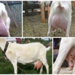
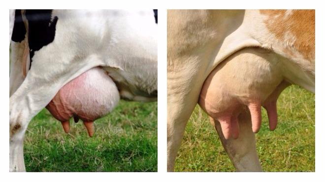
- Bath-shaped. The shape is typical for dairy breeds. It is deep, elongated, pushed forward, and looks oval from the side.
- Cup-shaped. The organ is round in shape and resembles a deep bowl in appearance.
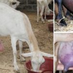
- Goat. It has pendulous rear and underdeveloped anterior nipples and a noticeable lateral furrow. Cows with such an udder are not suitable for machine milking; the shape is considered a developmental defect.
- Funnel-shaped. Tapering towards the bottom, with closely spaced nipples.
- Primitive. An underdeveloped organ with large nipples. It is formed if the heifer was poorly fed from birth.

The development of the mammary gland continues with the growth of the animal, but the udder of first-calf heifers especially increases in size during pregnancy. Then, over the years (up to about 6 calvings), the udder continues to grow, then the reverse process begins. Lactation directly depends on the state of the animal’s hormonal and reproductive systems.
How is milk produced?
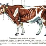
The lactation process is associated with the processes of digestion, metabolism, blood circulation, and respiration. The more glandular tissue the udder contains, the better it is supplied with blood, the higher the productivity of the animal.
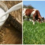
Lactation begins from the moment the cow first calves. Colostrum and milk are produced to feed the offspring. Milk is produced from products that enter the alveoli with blood, so a huge amount of blood passes through the udder; about 500 liters are needed to produce 1 liter of milk. The product in the udder of dairy cows is constantly formed; if the animal is not milked on time, its production decreases and then stops completely.
Over time, the cow develops a conditioned reflex: at the sound of the milking bowl, the sight of the mistress in certain clothes, grooming procedures, milk production increases. Acetic acid produced in the rumen is responsible for the fat content of milk; its production is regulated by the hormonal and nervous systems of the animal.
Udder hygiene
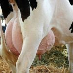
The udder performs the same functions as the female breast; you should care for it carefully. The organ must be inspected before each milking. The lobes should be symmetrical, the skin of the organ should be elastic and soft, and there should be no lumps or compactions in the tissues. Before milking, it is necessary to wash the mammary gland with warm water, use soap for severe contamination, then wipe the organ dry with a soft cloth.
Next, you should lubricate the udder with cream or ointment and give a light massage. To prevent debris from getting into the milk, not only the udder is washed, but also the stomach, sides, and hind legs.
The first drops of milk are milked into a separate bowl to clean the nipple canals. If the animal does not give milk well, repeat the massage during milking. Scratches, cracks, and abrasions of the udder must be treated with hydrogen peroxide, and after milking a layer of salicylic ointment is applied.The nipples do not have sebaceous and sweat glands, so the skin on them dries and cracks; the use of creams (Burenka, Lyubava, Zorka) relieves the problem.


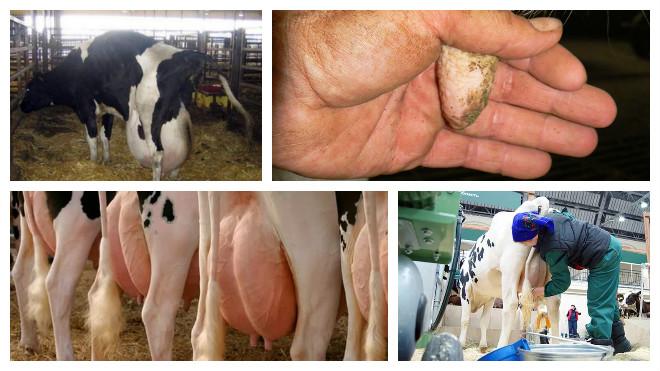
Possible diseases
Scratches, cracked nipples, and minor bruises can be treated on your own; if serious problems arise, you should consult a veterinarian.
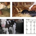
Mastitis
Inflammation usually occurs after calving. The udder or part of it becomes swollen, hot and hard to the touch. The cow is worried, loses appetite, and milk yield decreases. There may be traces of blood or pus in the milk. With serous mastitis, the milk takes on a bluish tint and flakes are visible in it.
In this case, the udder is gently massaged, the animal is milked by hand 5-6 times a day, and warming compresses are applied to the seal areas. In severe cases, antibiotic injections are prescribed.

Edema
If it is discovered after calving, no treatment is required. Frequent manual milking and light massage will get rid of the problem. Juicy foods should be excluded and salt should not be given. Lubricate the udder or its individual areas with Rigofen, bismuth-zinc ointment. In severe cases, calcium supplements by injection and caffeine are prescribed.
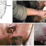
Smallpox
This is a serious infectious disease. The animal is isolated, the veterinary service is called, and quarantine measures are observed.
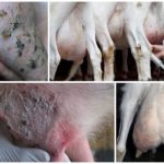
Furunculosis
Treatment requires frequent, 4-6 times a day, washing the udder with clean warm water and soap. Ichthyol ointment is applied to the boils, the skin area is treated with salicylic or camphor alcohol, and sprinkled with streptocide. Furunculosis occurs when there is cold and high humidity in the barn and non-compliance with sanitary standards.
Bruises
The bruise is lubricated with iodine, and a cold compress is applied for the first 2 days, then warming ointments are applied.You can use the “Rescuer” remedy for hematomas. It is applied in a thick layer. In severe cases, the area is opened and cleared of blood clots, and then the resulting wound is treated.
An antibiotic ointment (Levomikol, synthomycin emulsion) is applied. To ensure that compresses and napkins with ointment stay on the udder, a bandage is worn.
Insect bites
First, the sting is removed with tweezers. A cooling compress is applied to the bite site. Lubricate the bite site with Rigofen and Fenistil. On the recommendation of a veterinarian, antihistamines and drugs that support heart function are used.
Warts
If there are warts, apply salicylic ointment, or a mixture of salicylic and interferon ointment, to the affected areas 3-4 times a day. The course of treatment is 1-3 weeks.
To avoid mammary gland diseases, systematic tests for mastitis are carried out, animals are vaccinated, and creams and ointments are used to treat the udder. These drugs are sold in regular or veterinary pharmacies. Good nutrition, maintenance and careful care will allow animals to remain strong and healthy and ensure high milk yields.