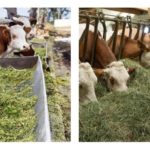
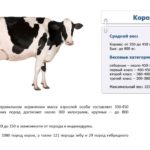
Every farmer should know the structure of the internal organs and the skeletal features of a cow in order to, if necessary, provide assistance to the animal on his own. Knowledge of cattle anatomy allows you to assess the full development of calves, identify fractures and internal injuries in animals, and keep the health of the herd under control. Anatomical knowledge is especially necessary for owners of small farms who do not have a veterinarian under their supervision.
- Head structure
- Scull
- Eyes
- Teeth
- Hearing aid
- How does the skeleton work?
- Spine
- Limbs
- Structure of internal organs and systems
- Muscles
- Nerves
- Respiratory organs
- Heart and blood vessels
- Digestive organs
- Stomach structure
- Urinary organs
- Reproductive system
- Udder structure
- Circulatory system
- Supplying the body with lymph
- Nerve endings
- Purpose of milk follicles
- Nipples
- Tail
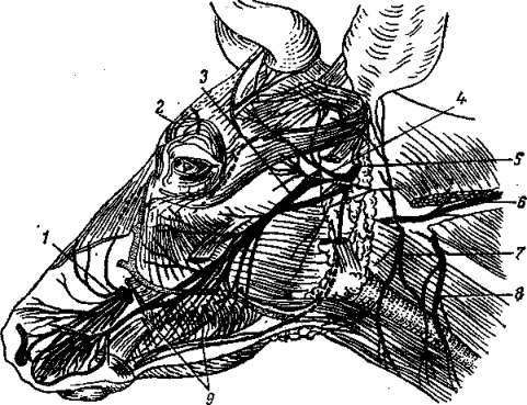
Head structure
A cow has a large head, consisting of a skull, eyes, ears, teeth, and nose.
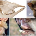
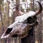
Scull
Cow skull divided into 2 sections: the first protects the brain, the second forms the muzzle with eye openings, nasal passages, jaws. In a calf, the sections are equal in volume; as the bull grows older, the facial section increases, but the brain section does not change.
The cranial skeleton of a cow is formed by 13 paired (symmetrically located on both sides) and 7 unpaired bones. Paired ones make up the crown, forehead and temples, unpaired ones make up the back of the head, sphenoid and interparietal parts. List of cow cranial bones:
- paired brain sections - frontal, parietal, temporal;
- paired facial - lacrimal, palatine, zygomatic, maxillary, mandibular, premaxillary, nasal, pterygoid, superior nasal concha, inferior concha;
- unpaired medulla - sphenoid, occipital, interparietal;
- unpaired facial - hyoid, ethmoid, vomer.
Eyes
The cow's visual organs are located symmetrically in the facial part of the skull. Cattle have monocular vision. The eyeball is located in the socket, it is round, slightly convex on the outside, covered with three membranes. Inside, the organ is divided into the vitreous body, the anterior and posterior lobes. Eyelashes – protection from mechanical influences. The tear glands secrete fluid that keeps the eyes moist. The iris of cattle is, in most cases, brown.
Teeth
Calves have 20 baby teeth. Adults have 32 teeth. The jaws of a cow are adapted for chewing plant foods. The incisors are long, directed forward, with sharp edges, growing from the lower jaw, designed for cutting grass.Chewing is carried out in a circular motion of the lower jaw.
Hearing aid
Cattle have good hearing. The cow's hearing organ consists of the outer, middle, and inner ears. The auricle is mobile, composed of muscle and cartilage tissue. The inside of the ears consists of the auditory ossicles and the eardrum.
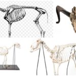
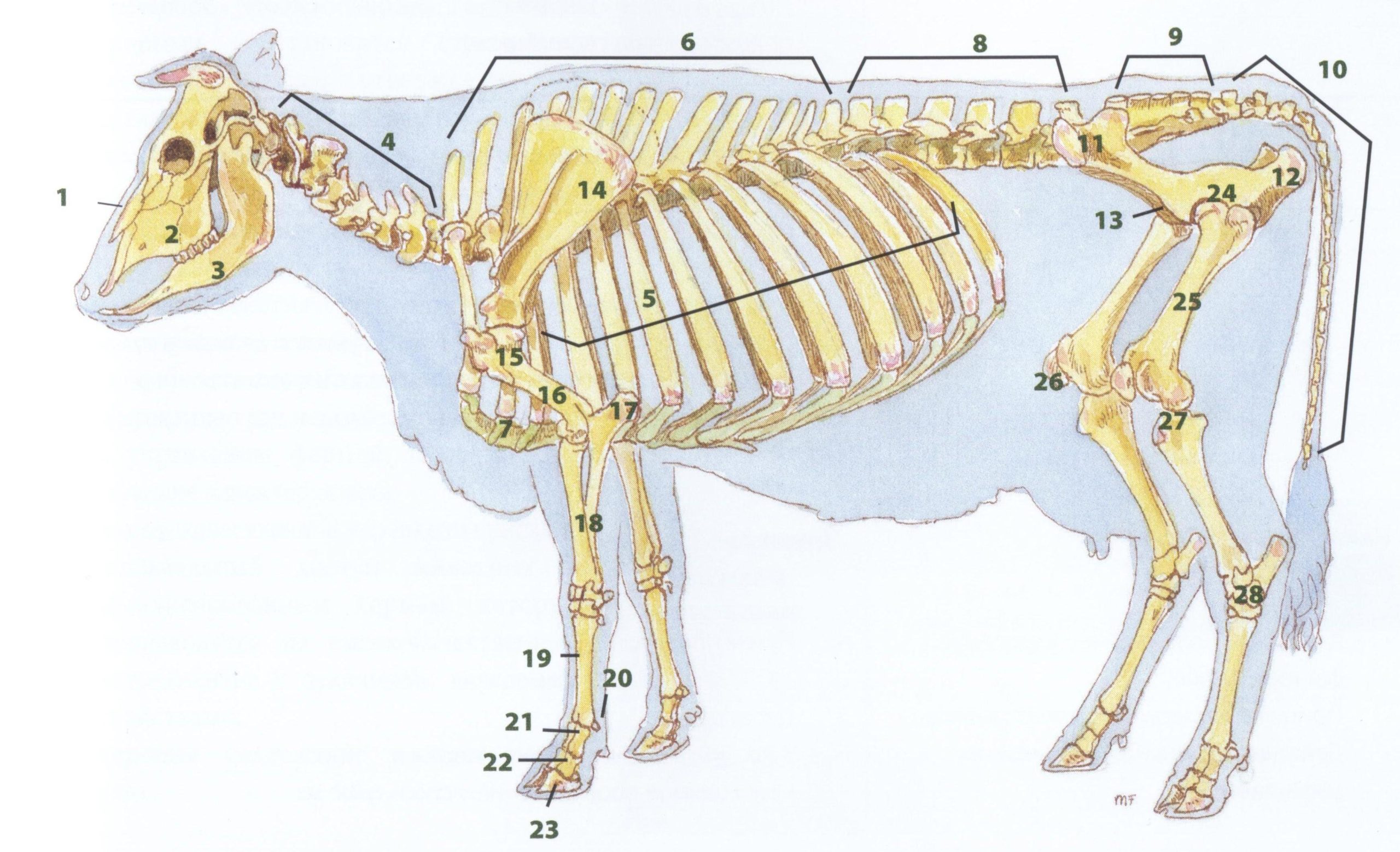
How does the skeleton work?
Cattle have a strong, heavy skeleton. Bulls have a more massive skeleton than females, which is due to greater muscle mass.
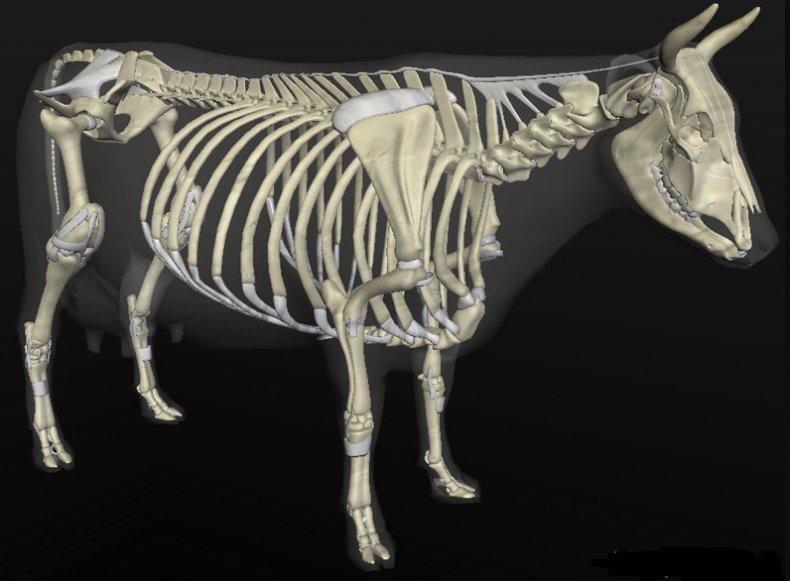
The skeleton of a cow consists of 2 parts:
- axial – cranium, spinal column, chest;
- peripheral – fore and hind limbs.
Spine
A cow has 50 vertebrae, the axial part of the skeleton includes:
- 7 cervical vertebrae;
- 13 breast;
- 6 lumbar;
- 5 sacral;
- 19 tail.
The cervical vertebrae are the most mobile, connecting the skull and sternum. Withers - 7th cervical vertebra. The thoracic skeleton is the least mobile; it is the basis for the attachment of the ribs. Ribs - 13 pairs of flat bones that form the rib cage, protecting the heart and lungs from injury. In a cow, 5 pairs of ribs are connected by cartilage, 8 pairs are free.
The description of the thoracic skeleton should be examined in more detail, since the anatomy of the costal plates is not the same. The front ribs are powerful and strong. The middle ones are widened towards the edge. The hind legs are short and curved. The last pair of ribs is attached only to the spine and does not reach the sternum.
Limbs
The skeleton of the forelimbs of cattle consists of the scapula, humerus, forearms, and hands. The hand is formed from the metacarpal, carpal, and phalangeal bones. The phalanges of the toes form the hooves. The skeleton of the forearm is formed by the ulna and radius bones. The radius bones of a cow are better developed than the ulnas.
The skeleton of the back of the body - pelvic bones, femurs, legs, feet. The femur is the largest bone in the cow skeleton.
Structure of internal organs and systems
The cow lives fully thanks to properly functioning internal organs and systems.
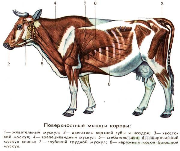
Muscles
When a calf is born, up to 80% of its body weight is musculoskeletal, which includes the skeleton and muscle tissue. In an adult cow, the skeleton and muscles make up about 60% of its weight.
The musculature of bulls includes 250 muscles. The full functioning of the body is ensured by the fact that the external muscular covering of the skeleton and internal smooth muscles form a functional complex.
In cross-section, the musculature of a cow consists of several main muscle groups:
- facial – regulate facial expressions, movements of the eyes, nostrils, lips;
- chewing – move the jaws;
- shoulder – move the shoulder skeleton;
- sternal – support the organs of the thoracic cavity, expand and move the chest during breathing;
- vertebrates – move the head, neck, spine, lumbar, pelvic, caudal sections of the skeleton;
- abdominal - support the abdominal organs, ensure defecation, urination, the functioning of the digestive tract, and uterine contractions.
Nerves
From the senses, signals travel along nerve fibers to the brain and are processed there. Brain impulses are sent to the senses and carry information about how to react to stimuli.
The nervous system of a cow is divided into several sections that have functional features:
- The brain is the basis of the central nervous system, controlling all life processes. A cow's brain weighs 550 g, is divided into equal hemispheres, and is covered with a membrane - the cortex.
- The spinal cord is a continuation of the central nervous system and is located in the canal of the vertebral skeleton. Reaches 1.8 m, controls unconditioned reflexes.
- Peripheral nerves are connectors of the brain with muscles, blood vessels, abdominal and secretory organs.
- Autonomic nerves are nodes that control external secretion, the functioning of the visual and respiratory organs, pelvic and abdominal organs, and smooth muscles.
Respiratory organs
The lungs of cattle are large, since the body of large animals requires a significant supply of oxygen. The lungs of a cow weigh 3500 g, a bull's - 4800 g. A cow's right lung is larger than the left. On the left side of the chest there is a large heart, which reduces the volume of the lung, and in some individuals almost divides it into two parts.
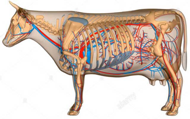
Heart and blood vessels
A cow has a four-chambered heart: 2 atria at the top, 2 ventricles at the bottom. Blood carries hormones and immune agents through vessels, and supplies nutrients, oxygen, and fluid to tissues and organs. Diagram of how a cow's heart works:
- When the heart muscle relaxes, the atria and ventricles fill with blood.
- The atria contract - a phase called systole. Blood flows into the ventricles.
- The atria relax. The valves separating them from the ventricles slam shut.
- The ventricles contract. During systole, blood is ejected from the left ventricle into the aorta, and from the right into the pulmonary artery.
- This is followed by diastole - relaxation of the organ, filling it with blood.
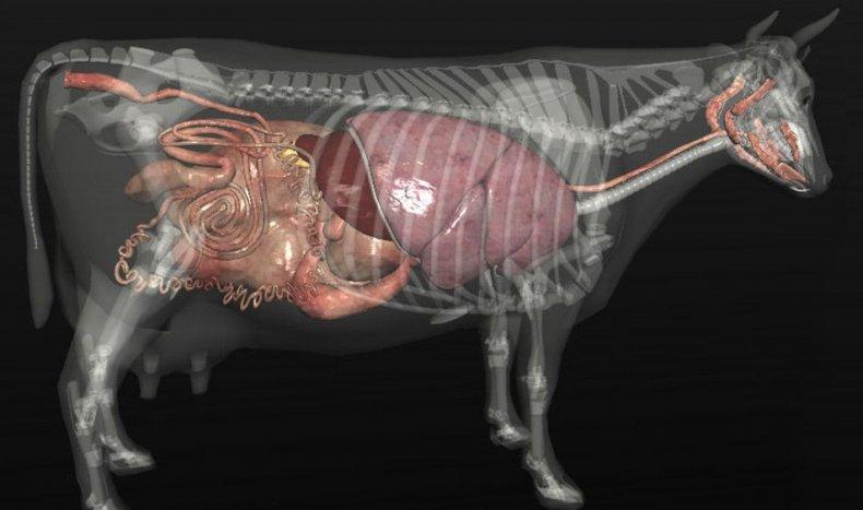
Digestive organs
Digestive system of a cow consists of several organs:
- Oral cavity. It chews food and secretes saliva.
- The esophagus is the tube through which chewed food moves into the stomach.
- The stomach is the organ for digesting and breaking down food particles.
- Pancreas. Located on the side of the stomach in the right hypochondrium. Produces digestive juice.
- Small intestine. Consists of duodenum, jejunum, ileum. It sucks nutrients from digested food.
- Colon. Consists of the cecum, colon, and rectum. Fermentation of the food mass occurs in it, the formation of feces, and its removal out through the anus.
The length of the cow's intestines is 63 m, which is 20 times the length of the body. Food that enters the digestive tract is digested within 2-3 days. A healthy cow excretes 20-40 kg of feces per day.
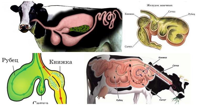
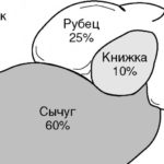
Stomach structure
Rough plant food is digested in the cow’s stomach, which has 4 sections:
- scar;
- mesh;
- book;
- abomasum.
A cow's rumen holds 200 liters. Here beneficial microflora breaks down fiber. The animal regurgitates the coarsest parts of the food so that they reenter the rumen and are thoroughly digested. Honeycomb structure mesh with a volume of 10 liters. Here the food mass remains for 2 days and is processed by microorganisms. Next, the food enters a book consisting of many thin plates. Here, liquid is absorbed within 5 hours. Digestion is completed in the rennet, which holds 10-15 liters, and the food mass is exposed to digestive juice.
Urinary organs
The excretory system of a cow consists of the kidneys, ureters, bladder and urethral canal.
The kidneys are a filtration organ. Purifying the blood from waste products, they produce 20 liters of urine per day. Urine is sent through the ureters to the bladder, where it accumulates to be released through the urethra.
Reproductive system
The genital organs of bulls are intended for the synthesis of sperm and fertilization of eggs:
- penis – organ of urination and removal of sperm;
- prepuce - the sheath of the outer edge of the penis;
- urethral canal;
- vas deferens - a channel for releasing sperm;
- spermatic cord - abdominal fold containing the vas deferens;
- testicles – organs of sperm synthesis and accumulation;
- The scrotum is a skin sac that contains the testicles.
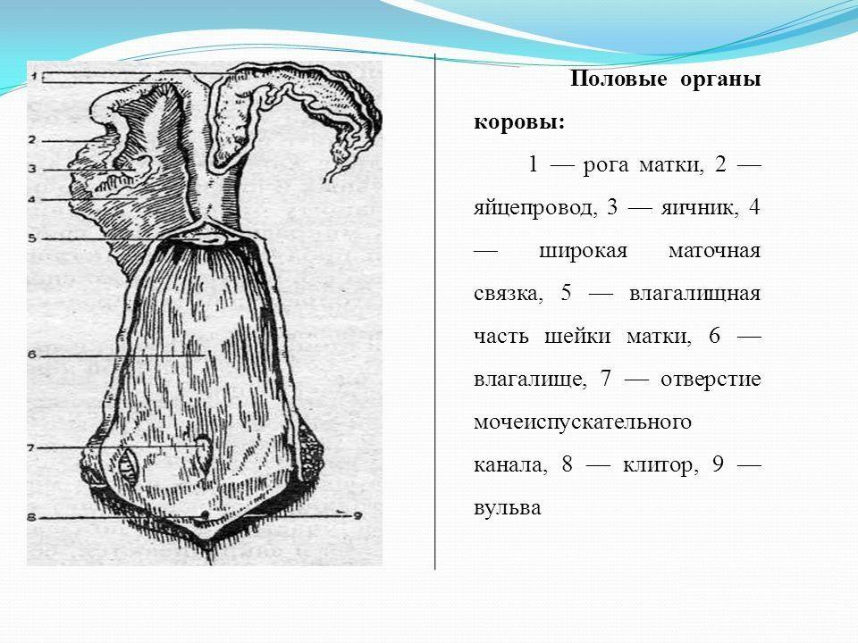
The female reproductive system is designed for bearing and giving birth to offspring:
- vagina;
- clitoris – amplifier of uterine contractions;
- labia;
- the uterus is a muscular organ that houses the developing embryo;
- fallopian tubes, through which the egg travels from the ovaries;
- Ovaries are egg storage organs.
Udder structure
The udder of a cow is divided into 4 parts. Each mammary gland ends with a nipple. That is, a cow has 4 teats.
Circulatory system
The mammary glands are abundantly entangled with blood capillaries carrying oxygen and nutrients.
Supplying the body with lymph
Separate from the blood capillaries, lymphatic vessels pass through the udder. They supply tissues with fluid and remove waste products.
There are lymph nodes on both sides of the udder. Their swelling signals the onset of mastitis.
Nerve endings
There are an abundance of nerve endings approaching the mammary glands. They transmit signals to the brain about the need for the synthesis and secretion of milk. Response signals from the brain make the cow worry and moo to tell the owner that the time for milking has come.
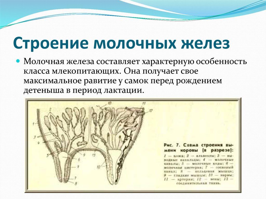
Purpose of milk follicles
The task of the follicles in the mammary glands is to excrete milk. The fluid accumulated in the milk tanks flows out through the nipple canals. The volume of follicles changes at different stages of a cow’s life - during estrus, pregnancy, lactation.
Nipples
The length of a cow's nipple is 8-10 cm, diameter is 3 cm. The nipple is not only a channel for the flow of milk, but also protects the mammary glands from external infection.It is divided into apical, main, cylindrical parts and body.
Tail
The vertebral skeleton ends with movable caudal vertebrae. The cow's tail is long, whipping, with a brush at the end, designed to brush blood-sucking insects from the body. Cows are strong, hardy animals with a strong skeleton and well-developed muscles. The health of livestock depends on the proper functioning of organs and systems, which must be maintained with proper care, maintenance and feeding.