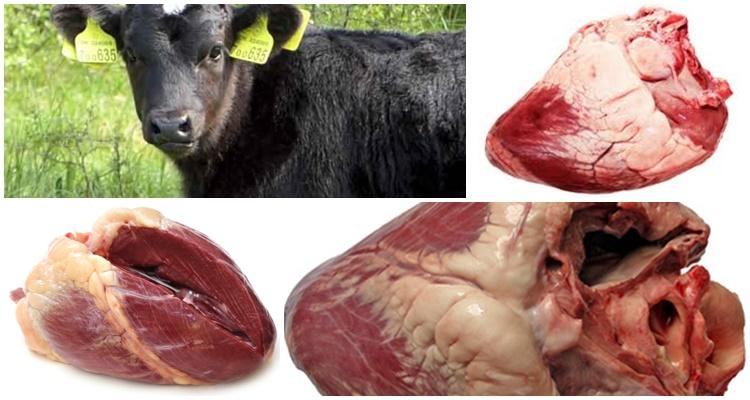
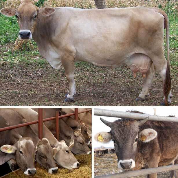
During the day, a healthy heart muscle of cattle pumps thousands of tons of blood. It is a hollow, continuously working, cone-shaped organ located between the 3rd and 6th ribs. The health of the body depends on the functional state of the cow’s heart; the organ supplies oxygen, nutrients, fluid to the tissues, supports metabolism and the full functioning of internal organs.
How does the heart apparatus work?
The cow's heart is chambered, the muscle tissues of the chambers contract with a certain rhythm, moving blood flows along a constant path: from the venous vessels to the atria, from there to the ventricles, then to the arteries. Continuity of movement and immutability of the blood path are ensured by valves. The work of the organ can be divided into three stages:
- compression (systole) – pushing out the contents of the heart cavity;
- blood promotion;
- relaxation (diastole) – filling of the cavity with blood.
In a healthy cow, the listed stages alternate with a clear frequency. When the ventricles work, the pressure inside them increases, the atrioventricular valves close, and the semilunar valves open later. As a result, blood leaves the heart. After the semilunar valves open, the blood flows more calmly, and therefore the myocardium begins to contract more slowly.
How does the cattle heart work?
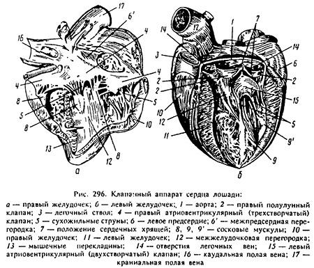
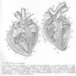
A cow's heart consists of 4 chambers: 2 atria located in the upper part, and 2 ventricles occupying the lower part of the organ. The interior is covered with endocardium. The upper and lower chambers are united by atrioventricular lumens.
Atria
The atria (atrium) occupy a small part of the upper half of the heart, separated from the ventricles on the outside by the coronary groove. The structure of the chambers is simple; their main element is the pectineal muscles, which, when contracting, push blood through.
The ventricles (ventriculus) are the main part of the organ in terms of volume. The ventricular chambers are not connected, they are longitudinally delimited by grooves. The connection between the atria and ventricles is provided by valves.
Valve apparatus
In the cow's heart, atrioventricular and semilunar valves function, opening and closing in accordance with the contractile work of the ventricles and atria. The right atrioventricular valve is tricuspid, the left is bicuspid. The anatomy of the atrioventricular lumens is such that during the functioning of the atria, the valves are pressed by blood to the ventricle. And when the ventricles begin to function, blood pressure raises the valves, forcing them to close the gaps. Semilunar valves, shaped like pockets, close the bases of the arteries.
How intensely the heart contracts depends on:
- meteorological conditions;
- age of the cow;
- physical condition of the body.
In a newborn calf, blood moves at a frequency of 140 pulsations per minute. In one-year-old cows, the indicator decreases to 100 pulsations, in adult cows – to 60.
Fibrous skeleton
Fibrous rings are adjacent to the aorta and two atrioventricular lumens. Over the years, the cartilage tissue covering these organ elements becomes thicker. Inside the rings are the left and right heart bones. In fact, fibrous formations are the skeletal basis of the heart, on which muscle tissue and valves are supported.
Circulation circles
Like all mammals, cows have two circulations:
- Big – systemic. The origin is the aorta, emerging from the left ventricle. The end is the venous vessels entering the right atrium.
- Small. The origin is the pulmonary artery, exiting the right ventricle. The end is the pulmonary vein, directed into the left atrium.
The structure of the heart and circulatory system ensures that it is impossible for venous (carrying carbon dioxide) and arterial (oxygen-saturated) blood to mix.
Vessels and nerves of the heart
Large vessels are connected by anastomoses - tiny capillaries. Anastomoses are:
- arterial - connecting two arteries;
- venous - two veins;
- arterial-venous - connecting artery and vein.
The functioning of the heart muscle is ensured by the autonomic nervous system. Sympathetic nerves approach the heart, stimulating muscle contractions. And the parasympathetic nerves weaken the contractile work of the heart.
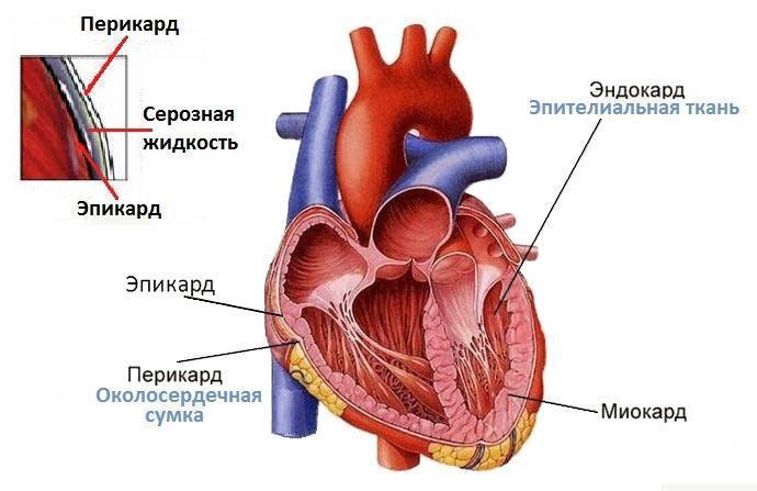
Pericardium (pericardium)
The cow's heart is surrounded by a film of connective tissue. Its task is to protect the heart from surrounding tissues, protect it from mechanical stress, and provide conditions for uninterrupted operation.
Layers of the heart wall
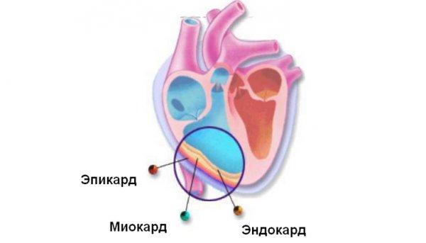
The walls of a cow's heart are made up of three types of tissue - endocardium, myocardium, and epicardium.
Endocardium
It lines the inside of the heart muscle and has unequal thickness in different parts of the organ. On the left it is denser, and is thinnest in the area of the tendon strings attached to the left atrioventricular valve. The endocardium of a cow consists of 4 layers:
- external – endothelium;
- subendothelial, consisting of loose connective tissue;
- muscular-elastic;
- muscular.
The fibrousness of the ventricular endocardium is less pronounced than that of the atria.
Myocardium
The muscle layer, in the thickness of which there are nerve fibers responsible for the contractile work of the heart.
Epicard
The outer lining of a cow's heart. Consists of two layers:
- external – mesothelium;
- internal, consisting of soft connective tissue.
Possible diseases
When the functioning of the heart muscle is disrupted, the entire body suffers: metabolism deteriorates, internal organs do not work correctly due to lack of oxygen and nutrients.The health and productivity of the cow are significantly reduced, so farmers should know what symptoms indicate heart pathologies that require immediate treatment.
The symptoms of myocardial fibrosis are as follows:
- swelling;
- rapid breathing;
- weakly audible pulse;
- tachycardia or arrhythmia;
- muting pulsation tones while listening.
Myocardial fibrosis in cows takes a long time to develop and manifests itself after several months. A sick cow should be kept in a warm barn, a high-quality and balanced diet should be selected for her, and she should be fed small portions several times a day. The veterinarian prescribes medications that improve blood circulation and suppress the development of the disease.
Myocarditis is an inflammatory process in the myocardium, leading to functional disorders of the heart. The inflamed organ has difficulty contracting. Most often, the disease occurs in cows that have suffered from intoxication or infection.
Symptoms of myocarditis in cattle:
- increased body temperature;
- rapid pulse;
- rapid or extraordinary contractions of the heart chambers;
- poor appetite;
- high blood pressure;
- rapid breathing;
- swelling;
- blue tint of the mucous membranes, skin around the nose and mouth.
Myocarditis is a serious disease that leads to disruption of the functional state of many internal organs. A sick cow should be kept warm and dry, fed in small portions, and given water heated to a comfortable temperature. The veterinarian identifies the cause of the pathology, prescribes medications that extinguish the inflammatory process and normalize the tone of the heart muscle.
Myocardosis – myocardial dystrophy. Often develops from untreated myocarditis.
Symptoms of myocardosis:
- weakening of the cow;
- failure of the rhythm of heart contractions;
- swelling;
- the cow's reluctance to eat food;
- a sharp drop in blood pressure;
- decreased muscle tone;
- bluish tint of the mucous membranes and skin around the mouth and nose;
- decreased skin tone.
The sick cow is taken to a warm, dry and quiet room. Provide quality food in small portions. The veterinarian identifies the cause of the pathology and prescribes medications that help stop degenerative processes in the myocardium.
Hydropericarditis is an accumulation of serous fluid inside the pericardium without an inflammatory process. Pericardial dropsy is provoked by either other heart diseases or chronic insufficiency of capillary blood circulation. Signs of pericardial hydrocele in cattle:
- swelling of the soft tissues of the jaws;
- weakening of the cow;
- fluctuations in blood pressure;
- decrease in milk yield.
The veterinarian prescribes medications for the underlying heart pathology that caused the dropsy. To remove cavity fluid, he recommends diaphoretic, diuretic and iodine-containing medications. A sick cow is well fed and given plenty of water.
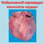
Pericarditis is an inflammation of the pericardium associated either with an infectious lesion or with injury to the heart sac. Cows that are poorly nourished are more likely to develop the disease because their metabolism is impaired.
Symptoms of pericarditis:
- weakening of the cow;
- body temperature either rising or falling;
- weak appetite;
- rapid breathing;
- decreased productivity;
- severe tachycardia;
- swelling of the chest, neck, abdomen;
- cow anxiety;
- the desire to adopt a position in which the chest is higher than the pelvis;
- weak pulsation, clear noises when listening.
With traumatic pericarditis, therapy is useless, the cow is sent to slaughter.In case of infectious pathology, the veterinarian prescribes antibiotics and drugs to restore heart function. The cow should be in a quiet place, eat light food, and have cold compresses placed on her chest.
The heart ensures the proper functioning of the cow's entire body. It is necessary to know the anatomy of the organ and the symptoms of pathologies in order to promptly identify life-threatening changes in the animal and begin treatment.

